Introduction
ARMD is a medical condition which usually affects older adults and causes a loss of vision in the centre of the visual field due to degeneration of receptor cells in the macula (the central area of the retina). It is the major cause of blindness and visual impairment in older adults (>50 years).
Macular degeneration can make it difficult or impossible to read or recognize faces, although enough peripheral vision remains to allow other activities.
Types of ARMD
Age-related macular degeneration begins with characteristic yellow deposits (related to deposits of cholesterol) in the macula. No medical or surgical treatment is available for this condition. Vitamin supplements with high doses of lutein and zeaxanthin (both antioxidants) have been shown to slow the progression of the condition.
Caused by abnormal blood vessel growth in the eye, it can lead to bleeding, leaking and scarring causing irreversible damage to the photoreceptors and rapid vision loss if left untreated.
It can be treated with laser coagulation, and with medication sometimes the condition reverses the growth of blood vessels. Only about 10% of patients suffering from macular degeneration have the wet type.
Symptoms of Macular Degeneration
Since the condition is not painful, it may go unnoticed for some time. Symptoms of macular degeneration include:
- Blurred vision: Those with non-exudative (dry) macular degeneration may be asymptomatic or notice a gradual loss of central vision, whereas those with exudative (wet) macular degeneration often notice a rapid onset of vision loss.
- Central missing areas of vision, also called scotomas.
- In certain cases, patients may complain that straight lines appear wavy. This form of distorted vision is called metamorphopsia.
- Patients may have trouble differentiating between colours and have low contrast sensitivity.
- Patients may complain about slow recovery of visual function after exposure to bright light.
Causes
Approximately 10% of patients in the age group of 66 to 74 years have macular degeneration. The prevalence increases to 30% in patients of the age group 75 to 85 years of age.
The lifetime risk of developing late-stage macular degeneration is 50% for people that have a relative with macular degeneration, versus 12% for people that do not have relatives with macular degeneration.
High cholesterol, obesity, high fat intake is associated with an increased risk of AMD in both women and men. A diet that derives closer to 20-25% of total food energy from fat is probably healthier.
Evidence is conflicting as to whether exposure to sunlight contributes to the development of macular degeneration. A recent study on 446 subjects found it does not. Other research, however, has shown high-energy visible light may contribute to AMD.
Smoking tobacco increases the risk of AMD by two to three times than that of someone who has never smoked, and may be the most important modifiable factor in its prevention. Cigarette smoking is likely to have toxic effects on the retina.
Does Macular Degeneration Lead To Total Blindness?
In almost all cases, some vision remains. The area of the macula comprises only about 2.1% of the retina, and the remaining 97.9% (the peripheral field) remains unaffected by the disease. Interestingly, even though the macula provides such a small fraction of the visual field, almost half of the visual cortex is devoted to processing macular information.
Diagnosis
There are various tests and investigations to diagnose the condition:
- The Amsler grid test is one of the simplest and most effective methods for patients to monitor the health of their maculae. The Amsler grid is, in essence, a pattern of intersecting lines (identical to graph paper) with a black dot in the middle. The central black dot is used for fixation (a place for the eye to focus). With normal vision, all lines surrounding the black dot will look straight and evenly spaced, with no missing or odd-looking areas when fixating on the grid’s central black dot. In macular degeneration, the lines can look bent, distorted and/or missing.
- Fluorescein angiography allows for the identification and localization of abnormal vascular processes.
- Optical coherence tomography is now used in the diagnosis and the follow-up evaluation of the response to treatment by using either Avastin or Lucentis, which are injected into the vitreous humor of the eye at various intervals.
Treatment
Drugs approved for some variety of macular degeneration include Ranibizumab and Bevacizumab.
Treatments for wet ARMD:
- Certain injectable drugs containing anti-angiogenics or anti-VEGF agents can cause regression of the abnormal blood vessels and improve vision when injected directly into the vitreous humor of the eye. The injections must be repeated monthly or bimonthly. Several anti-angiogenic drugs have been approved for use in the eye by the U.S. Food and Drug Administration and regulatory agencies in other countries.
- Photodynamic therapy has also been used to treat wet AMD.
- Some evidence supports a reduction in the risk of AMD with increasing intake of two carotenoids, lutein and zeaxanthin.
- Consuming omega-3 fatty acids (docosahexaenoic acid and eicosapentaenoic acid) has been correlated with a reduced progression of early AMD, in conjunction with low glycemic index foods.
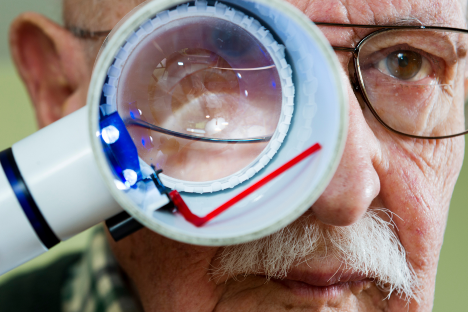
- Adaptive devices can help people affected with ARMD use their peripheral vision better and compensate for their lack of central vision. These include magnifying glasses, special eyeglass lenses, computer screen readers, and TV systems that enlarge reading material.
- Computer screen readers such as JAWS or Thunder that work with standard Windows computers can also help.
- Video cameras can be fed into standard or special-purpose computer monitors, and the image can be zoomed in and magnified. These systems often include a movable table to move the written material.